The problem the cochlea solves
The ear faces a translation problem. Sound in the air is pressure variation, waves of compression and rarefaction. The brain operates on electrochemical signals in neurons. Bridging these two entirely different forms of information requires several mechanical and cellular steps. The cochlea is where the critical translation happens.
Understanding how the cochlea works is relevant to tinnitus because most tinnitus originates in or near the cochlea, and because the cochlea’s specific design helps explain why certain kinds of damage produce ringing at particular pitches.
The path from outside to inside
Before a sound reaches the cochlea, it travels through two mechanical stages.
First, the outer ear, the visible pinna and the ear canal, channels sound waves to the eardrum. The shape of the pinna helps localize sounds and adds some amplification in the frequencies important for speech.
Second, the middle ear converts the air-pressure vibrations at the eardrum into fluid-pressure vibrations in the inner ear. Three tiny bones, the ossicles, the malleus, incus, and stapes, form a mechanical chain from the eardrum to the oval window, a membrane-covered opening on the cochlear wall. The ossicles act as a lever and an impedance-matching transformer, because transferring energy from light air into dense fluid would normally involve huge losses. The middle ear’s mechanical arrangement reduces those losses significantly.
The cochlea’s physical structure
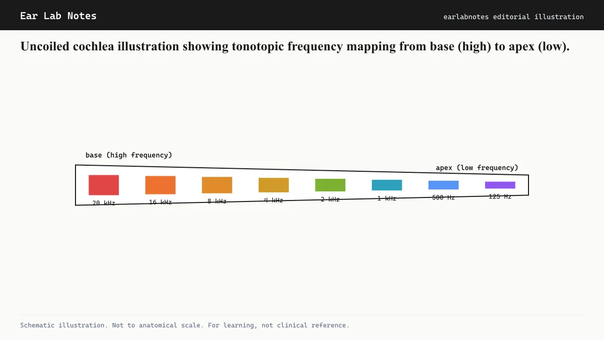
The cochlea is a hollow bony structure coiled into roughly two and a half turns. If you imagined stretching it out into a straight tube, it would be about 35 millimeters long, slightly longer than a large paperclip.
Inside, the cochlea is divided longitudinally into three fluid-filled chambers by membranes:
- The scala vestibuli (upper chamber) connects to the oval window where the stapes presses.
- The scala tympani (lower chamber) connects to the round window, another membrane-covered opening that accommodates the fluid displacement the oval window creates.
- The scala media (middle chamber, also called the cochlear duct) runs between the other two and contains the organ of Corti, where hair cells live.
The scala vestibuli and scala tympani are connected at the apex of the cochlea through a small gap called the helicotrema. They contain perilymph. The scala media contains endolymph, a chemically unusual fluid that is high in potassium and low in sodium, maintained by the stria vascularis through active ion pumping.
The basilar membrane and the traveling wave
Running along the length of the cochlear duct is the basilar membrane. This membrane changes in physical properties from one end to the other. At the base (near the oval window), it is narrow and stiff. At the apex (at the far end of the coil), it is wide and floppy.
When the stapes presses on the oval window, it creates a pressure wave in the cochlear fluid. This wave travels along the basilar membrane as a traveling wave, a rolling displacement rather than a simple vibration. The key property of the traveling wave is that different frequencies cause maximum displacement at different locations along the membrane.
High-frequency sounds (high pitches) create maximum displacement near the stiff base of the cochlea. Low-frequency sounds (low pitches) create maximum displacement near the floppy apex. This physical frequency-to-place mapping is called the tonotopic map, and it is the cochlea’s fundamental organizational principle.
NIDCD educational materials on hearing describe this property as the basis for the inner ear’s ability to distinguish pitch, because the brain receives spatial information, which location along the cochlea is responding most strongly, and interprets it as pitch.
The organ of Corti
The organ of Corti sits on top of the basilar membrane and contains the hair cells that perform the actual signal transduction, the conversion of mechanical motion into electrical signals.
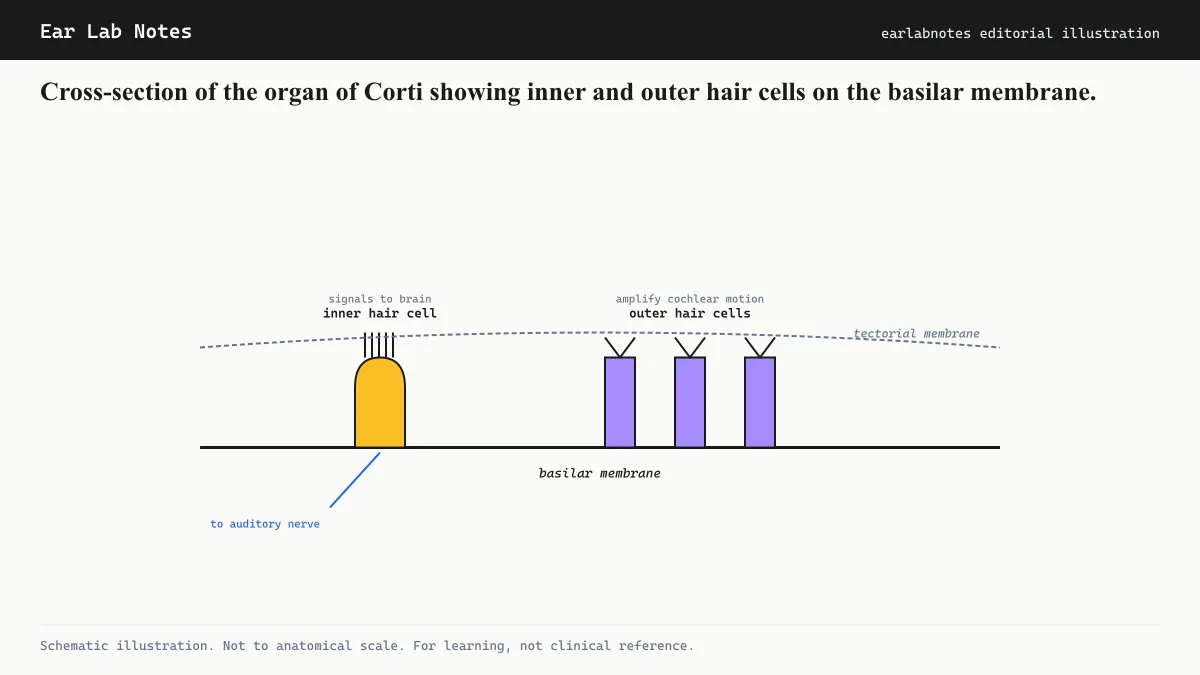
The organ of Corti contains two types of hair cells:
Inner hair cells form a single row running the full length of the cochlea, approximately 3,500 cells in total. These are the primary sensory cells. When the basilar membrane displaces, it bends tiny hair bundles (stereocilia) at the top of these cells. The bending opens ion channels, potassium flows in from the endolymph, the cell depolarizes, and neurotransmitter is released onto waiting auditory nerve fibers. The auditory nerve then carries the signal toward the brainstem and ultimately the auditory cortex.
Outer hair cells number approximately 12,000 and arrange in three rows. These cells do something unexpected: they change their length in response to electrical signals, contracting and elongating at the frequency of the sound wave. This active motility amplifies the mechanical response of the basilar membrane, making the cochlea far more sensitive and frequency-selective than it would be if it were passive. Outer hair cells are the cochlea’s built-in amplifier.
Stereocilia: the mechanical detector
Each hair cell has a bundle of stereocilia, tiny projections arranged by height. The tallest stereocilia in each bundle are connected by delicate tip links to shorter neighbors. When the bundle deflects, the tip links pull on mechanically gated ion channels and open them. The precision required here is extraordinary: displacements as small as a fraction of the diameter of a hydrogen atom can be detected by healthy hair cells. This sensitivity is what makes whispered speech audible.
Tonotopy and tinnitus
Because the cochlea maps frequency to position, damage to a specific region of the cochlea affects hearing at specific frequencies. Noise exposure at high volumes tends to damage the base of the cochlea first, because the base responds to the frequencies most prominent in noise, particularly around 3 to 6 kHz. This is why noise-induced hearing loss characteristically produces a notch in the audiogram at 4 kHz.
When hair cells at a particular location are damaged or destroyed, the auditory nerve fibers that connected to them lose their normal input. Research reviewed by NIDCD suggests that these deafferented nerve fibers may become spontaneously active, and that the brain regions receiving their input may reorganize in ways that generate or amplify phantom signals at the frequency the damaged cochlear region used to process. This is one of the leading models for why tinnitus pitch often corresponds to the frequency of greatest hearing loss.
The regeneration problem
Mammalian cochlear hair cells do not regenerate after damage. Unlike skin or liver cells, hair cells that die from noise, aging, or other injury are not replaced. Some non-mammalian vertebrates, including birds, can regenerate cochlear hair cells, which has driven substantial research into whether the regenerative pathways could be activated in humans. As of current knowledge, no such treatment is available, which is why hearing protection and noise limitation remain the only fully reliable strategies against cochlear hair cell loss.
If symptoms persist or change, see an audiologist or physician.
Related notes
Watch
Journey of Sound to the Brain
Source: NIDCD / NIH on YouTube
Transcript / summary
Frequently asked
Questions readers ask
- How does the cochlea convert sound to nerve signals?
- Sound waves move the eardrum, which moves the ossicles, which press on the oval window of the cochlea. This creates a fluid wave in the cochlea that displaces the basilar membrane. Hair cells on the basilar membrane detect the displacement and release neurotransmitters that trigger signals in the auditory nerve.
- How many hair cells does the human cochlea have?
- The human cochlea contains roughly 16,000 hair cells arranged along the organ of Corti. About 3,500 are inner hair cells responsible for signaling to the brain; around 12,000 are outer hair cells responsible for amplifying the cochlear response.
- Why does the cochlea look like a snail shell?
- The cochlea is coiled into roughly 2.5 turns, largely as a space-efficient solution. If uncoiled, the human cochlea would be approximately 35 millimeters long. Researchers also note that the coiled shape may help tune the mechanical properties of the basilar membrane.
- Can cochlear hair cells regenerate if damaged?
- In humans, cochlear hair cells do not regenerate after damage. This is a fundamental limitation of the human auditory system, and it is why noise-induced hearing loss and the tinnitus associated with it can be permanent. Some species of birds can regenerate cochlear hair cells, which has driven significant research interest.
- What is the tonotopic map and why does it matter for tinnitus?
- The tonotopic map is the cochlea's built-in frequency organization, where each location along the basilar membrane responds best to a specific frequency. When hair cells at a particular location are damaged, the brain region that receives signals from that location may generate phantom activity at the corresponding frequency, which is one explanation for why tinnitus pitch often correlates with the frequency of greatest hearing loss.
Primary sources
Where this comes from
- ◆ How Do We Hear? · NIH/NIDCD
- ◆ Noise-Induced Hearing Loss · NIH/NIDCD
- ◆ Ear Anatomy · Mayo Clinic
- ◆ How the Ear Works · NHS UK
Educational use only.
If your symptoms persist or change, see a licensed audiologist or otolaryngologist. Sudden hearing loss is a medical emergency, see a clinician within 72 hours.
TEL—N015 · The Ear Lab · earlabs.app