Two cell types, two jobs
The organ of Corti contains roughly 16,000 hair cells, but not all hair cells are the same. They divide into two functionally distinct populations with complementary roles. Understanding the difference explains a great deal about how noise causes tinnitus and hearing loss.
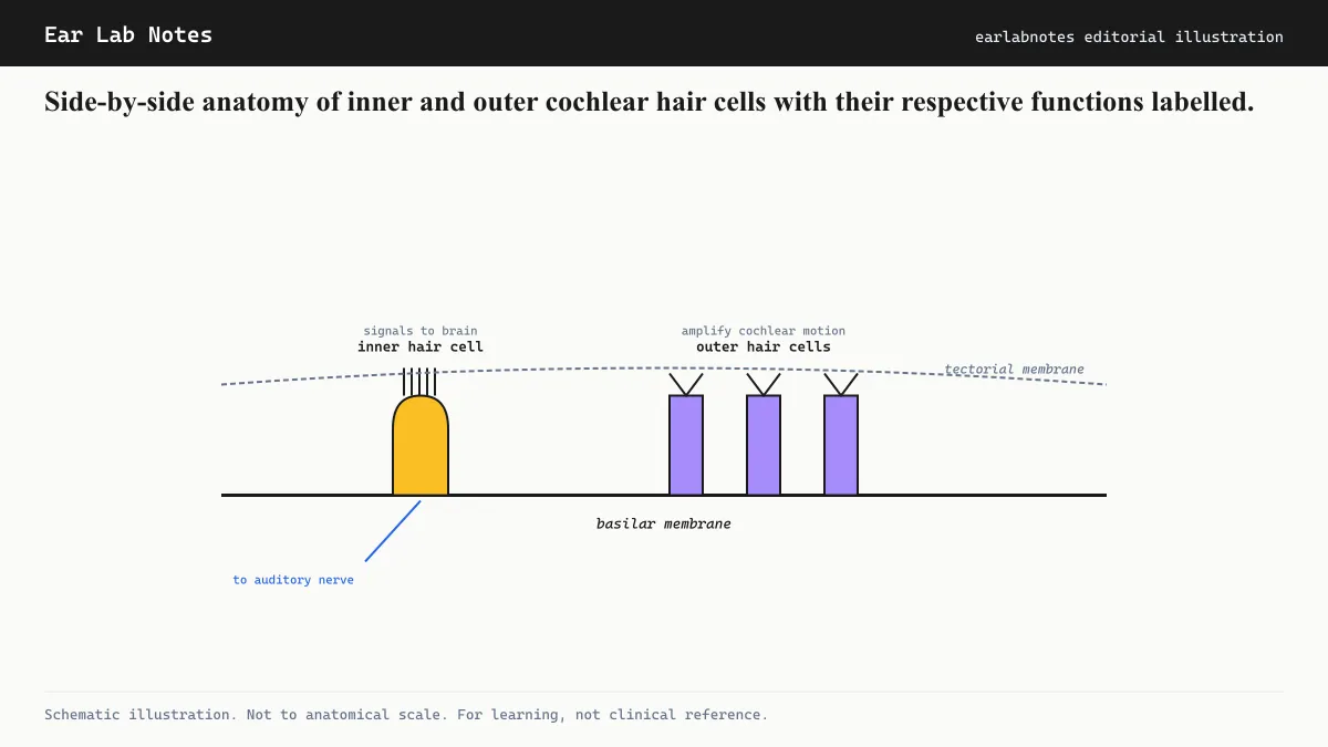
Inner hair cells are the sensory neurons in the physiological sense. There is one row of them running the full length of the cochlea, about 3,500 cells in total. Their job is to detect basilar membrane displacement and convert it into neurotransmitter release, triggering signals in the auditory nerve. Approximately 95 percent of all auditory nerve fibers connect to inner hair cells.
Outer hair cells are mechanics. There are three rows of them, roughly 12,000 cells. They detect basilar membrane movement, but instead of primarily sending signals to the brain, they change their own length in response to electrical signals. This active length change amplifies and sharpens the basilar membrane’s response to sound. Without outer hair cells, the cochlea would be roughly 40 to 60 dB less sensitive and would lose much of its ability to resolve nearby frequencies.
Why the shapes are different
Inner hair cells are flask-shaped: a wide cell body, a rounded bottom, and a flat top supporting stereocilia arranged in a relatively straight row. Their innervation is rich on the afferent (toward the brain) side and sparse on the efferent (from the brain) side.
Outer hair cells are cylindrical: elongated tubes with contractile elements built into their lateral walls. The protein responsible for their motility is called prestin, and it is one of the fastest motor proteins known in biology. Prestin changes shape in microseconds in response to changes in membrane voltage. This allows outer hair cells to track and amplify vibrations at frequencies up to 20,000 Hz in humans. Their innervation is the reverse of inner hair cells: rich on the efferent side, sparse on the afferent side.
This difference in innervation pattern reflects their roles. Inner hair cells report to the brain. Outer hair cells receive instructions from the brain (via the olivocochlear bundle), which allows the auditory system to modulate its own sensitivity, turning down gain in loud environments and potentially contributing to selective attention.
The stereocilia bundle
Both cell types have stereocilia, the mechanically gated hair bundles at their tops, but the arrangement differs.
In both types, stereocilia are arranged by height in a staircase pattern, with the tallest at one edge. Adjacent stereocilia are linked by tip links, delicate molecular springs made of cadherin proteins. When the bundle deflects toward the tallest stereocilia, tip links pull on mechanically gated ion channels and open them. Potassium ions from the surrounding endolymph rush in, depolarizing the cell.
Outer hair cell stereocilia bundles are arranged in a V or W shape, pointing outward. The tips of the outer hair cell tallest stereocilia are embedded in the overlying tectorial membrane. When the basilar membrane moves, the tectorial membrane shears across the hair bundle, producing a defined deflection. This coupling is part of what makes outer hair cells sensitive amplifiers.
Inner hair cell stereocilia are not embedded in the tectorial membrane. They are deflected by the fluid motion that accompanies basilar membrane displacement, making them somewhat less sensitive per deflection but still exquisitely capable of signaling at low sound levels when outer hair cell amplification is intact.
Why noise hits outer hair cells first
Noise-induced damage is not random. Outer hair cells at the base of the cochlea, which process high frequencies, are the most vulnerable. Several factors contribute:
Higher metabolic demand. Outer hair cells are continuously active, even in quiet environments. Their prestin motors run all the time, consuming ATP at a high rate. Metabolic stress from noise exposure, which includes increased reactive oxygen species, overwhelms outer hair cells before reaching inner hair cells.
Mechanical position. Outer hair cells sit on the outer edge of the organ of Corti and are more exposed to the largest basilar membrane displacements for high-frequency sounds.
The 4 kHz vulnerability. NIOSH noise exposure data consistently shows that the 4 kHz region of the cochlea suffers the most damage from typical occupational and recreational noise. The outer hair cells in this region show the classic noise notch pattern on audiograms before inner hair cell damage becomes apparent.
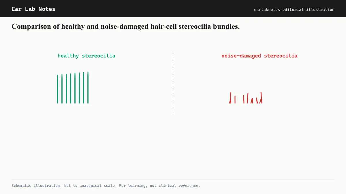
When outer hair cells are damaged, their stereocilia fuse, collapse, or disappear. The organ of Corti loses its amplification at the affected frequencies. Hearing thresholds rise because soft sounds are no longer amplified to the level required to drive inner hair cells. Frequency resolution degrades because the sharpness of the traveling wave peak, which outer hair cells produce, is reduced.
When inner hair cells and synapses are involved
For a long time, the dominant model was that detectable hearing loss required outer hair cell damage. More recently, research has established that the synapses connecting inner hair cells to auditory nerve fibers can be silently damaged before any outer hair cell loss appears and before audiometric thresholds shift. This is called cochlear synaptopathy or hidden hearing loss.
In cochlear synaptopathy, the inner hair cells themselves survive, but they lose some of the auditory nerve contacts. This reduces the total information capacity of the auditory nerve without changing hearing thresholds, because enough synapses remain to detect threshold-level sounds. The damage shows up instead in tasks that require high temporal precision or the ability to process sounds in noisy environments, where the reduced nerve fiber population cannot handle the signal complexity.
NIDCD-affiliated researchers have proposed that cochlear synaptopathy may be a source of tinnitus even in people whose audiograms look normal, which would help explain why some people develop tinnitus without apparent hearing loss on standard tests.
The non-regenerative constraint
A central fact about cochlear hair cells in humans is that they do not regenerate. Hair cells that die from noise, aging, or ototoxic exposure are not replaced. The approximately 16,000 cells present at birth are, in principle, all that will ever be available. Some non-mammalian species regenerate cochlear hair cells after damage, which has made understanding the molecular mechanisms of non-mammalian regeneration a significant research priority. As of current knowledge, no clinically available treatment can restore lost human cochlear hair cells.
This is why hearing protection, limiting noise exposure to levels below NIOSH thresholds, wearing appropriate ear protection in loud environments, and managing sound dose over time, remains the most reliable approach to preserving the hair cells that support hearing and reduce tinnitus risk.
If symptoms persist or change, see an audiologist or physician.
Related notes
Frequently asked
Questions readers ask
- What is the difference between inner and outer hair cells?
- Inner hair cells are the actual sensory cells that send signals to the auditory nerve and the brain. Outer hair cells are mechanical amplifiers that boost basilar membrane movement and sharpen the cochlea's frequency selectivity. The two types have very different shapes, innervation patterns, and vulnerabilities.
- Which hair cells are damaged by noise first?
- Outer hair cells are generally the first to be damaged by excessive noise exposure. They are more metabolically active and sit in a more mechanically stressed position on the basilar membrane. Inner hair cells tend to be more resilient and may survive moderate noise exposures that have already destroyed adjacent outer hair cells.
- Can you lose outer hair cells and still hear normally on a standard test?
- Standard audiograms measure hearing thresholds, the softest sound you can detect. Outer hair cells amplify soft sounds and sharpen tuning. If enough outer hair cells are lost, hearing thresholds do rise. However, early or scattered outer hair cell loss may not shift thresholds detectably, even as other hearing abilities, such as understanding speech in noise, are already degraded.
- Why does tinnitus pitch often match the frequency where hair cells are damaged?
- The cochlea maps frequency to location. When hair cells at a specific location are damaged, the auditory cortex region that processed signals from that location may generate spontaneous activity. The brain interprets that activity as sound at the corresponding frequency. This is why audiologists find tinnitus pitch often aligns with the edge of hearing loss on an audiogram.
- Do damaged hair cells recover?
- In humans, cochlear hair cells do not regenerate after damage. Some recovery of hearing threshold is possible in the hours to days after a single noise exposure if the damage was not complete, partly because of temporary metabolic disruption that can resolve. Permanent hair cell loss does not recover.
Primary sources
Where this comes from
- ◆ Noise-Induced Hearing Loss · NIH/NIDCD
- ◆ How Do We Hear? · NIH/NIDCD
- ◆ Noise and Hearing Loss Prevention · NIOSH/CDC
- ◆ Hearing Loss · NHS UK
Educational use only.
If your symptoms persist or change, see a licensed audiologist or otolaryngologist. Sudden hearing loss is a medical emergency, see a clinician within 72 hours.
TEL—N027 · The Ear Lab · earlabs.app